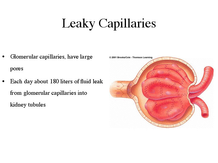

From Bowman's capsule, the urinary filtrate will move down the nephron tubule, becoming increasingly modified as it progresses. Before urine is ready to be excreted, it passes through 1) the proximal convoluted tubule, 2) the loop of Henle, 3) the distal tubule, and 4) the collecting ducts. The first step in modifying the urinary filtrate occurs in the proximal convoluted tubule. In this region, active transport pumps glucose, amino acids, sodium, and proteins back out of the filtrate. Water follows these by osmosis, concentrating the urine and reducing the volume of filtrate. This step conserves necessary metabolites that would otherwise be wasted in urine at the same time that it concentrates the urinary filtrate.
From the proximal tubule, the filtrate passes to the loop of Henle. While the glomerulus and Bowman's capsule of each nephron are located in the outer region of the kidney (the cortex), the loop of Henle dips down into the inner kidney region called the medulla. The medulla has a very high concentration of extracellular sodium. As the filtrate passes down the loop of Henle, water is drawn out of the filtrate due to osmosis, passing from the low ion concentration in the filtrate to the high ionic strength of the extracellular fluid in the medulla. When the filtrate passes back up the loop of Henle, sodium is pumped out into the medulla. These two steps further reduce the volume of the urinary filtrate, drawing out the water along with the sodium, and help to preserve the high concentration of sodium in the medulla.
After passing through the distal tubule, the filtrate must pass through the collecting duct before passing out to the ureter and the urinary bladder. The collecting duct passes back down through the high ionic strength medulla. To make concentrated or dilute urine, the hormone ADH (anti-diuretic hormone) regulates the permeability of the collecting duct walls.
This animation (Audio - Important) describes the structure of the kidney.
This animation (Audio - Important) describes the structure of the nephron.