Organisms make nutrients available to cells through the process of absorption. But these nutrients,
along with gases and wastes, must also be transported throughout these organisms' bodies in order
for them to survive. The process by which this transportation is accomplished is known as circulation.
The circulatory system is an elaborate maze of
arteries, veins, and capillaries. This is supplemented by the lymphatic system, which transports
lymph and recaptures fluid from the extracellular spaces to retum it to the blood.
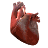
The human heart is a four-chambered pump, with two collecting
chambers called atria and two pumping chambers called ventricles. This organ is central to the
smooth functioning of the circulatory system, as it is the heart that keeps blood, the circulatory
fluid, moving throughout the body. The
right ventricle pumps deoxygenated blood to the
lungs through the pulmonary artery. Oxygenated blood retums through the pulmonary vein to the left
atrium. From there it passes to the left ventricle and is pumped through the aorta and arteries to
the rest of the body.
This animation (Audio - Important) describes the structure of
the heart.
The arteries carry blood from the heart to the tissues of the body. They repeatedly branch into
smaller arteries (arterioles), eventually supplying blood to the tissues via the capillaries.
Arteries are thick-walled, muscular, and elastic. They conduct blood at high pressure and have a
pulse (periodic surges of blood from the heart). Arterial blood is oxygenated (except for the
pulmonary artery, which carries deoxygenated blood from tissues to the lungs to renew the oxygen
supply).
Veins, on the other hand, carry blood back to the heart from the capillaries. Veins are relatively
thin-walled, conduct at low pressure (because
they are at some distance from the pumping heart), and contain many
valves to prevent backflow. Veins have no pulse. They usually carry dark
red, deoxygenated blood (except for the pulmonary vein, which carries
recently oxygenated blood from the lungs).
This animation (Audio - Important) describes what happens when the doctor checks your
blood pressure.
This information in this animation (Audio - Important) will not be on the final, but it will give
you information on the location and function of important arteries and veins
in your body.
Capillaries
Capillaries are thin-walled vessels that are very small in diameter. Their walls are only one
endothelial cell thick. Capillaries, not arteries or veins, permit exchange of materials between
the blood and the body's cells. Intercellular fluid, which contains plasma with digested
nutrients, enzymes, and hormones, seeps from capillaries through diffusion and bathes the cells.
Some of the fluid that enters tissues passes directly back into the blood, and the rest can
circulate back in the lymphatic system.
This animation (Audio - Important) describes the function of
capillaries.
These vessels are a separate system of tubes semi-independent of the blood system. This system
carries extracellular fluid (at this stage known as lymph) from one lymph node to the next at very
low pressure. The lymph nodes are responsible for
filtering lymph to rid it of foreign particles. The lymph
system ultimately returns lymph to the blood system via the largest lymph vessel, the thoracic
duct, which empties lymph back into circulation shortly before it enters the heart.
Circulation would not be possible without blood. One of blood's most vital
components is plasma, the liquid part of blood that contains dissolved nutrient wastes, proteins,
hormones, and fibrinogen. Several vital solid materials are suspended in plasma and transported
throughout the body, such as red and white blood cells, and platelets.
Red Blood Cells - These are the most common cells found in blood, and their primary function is to
transport oxygen. After they are formed in the bone marrow, red blood cells (erythrocytes) lose
their nuclei and become biconcave discs. They live for about four months. At the end of this period,
they are destroyed in the spleen. Red blood cells contain hemoglobin (the red
pigment containing iron), which unites with oxygen to form oxyhemoglobin. It is in this form that
oxygen is carried in the blood. In the tissues, the
partial pressure of oxygen is low and hemoglobin releases oxygen.
White Blood Cells - White blood cells are generally used for protective and defensive functions in
the body. White blood cells include phagocytes, which engulf bacteria with amoeboid motion, and
various types of lymphocytes, such as B and T cells which are involved in the immune response. B
cells produce antibodies, or immunoglobins, which are secreted proteins specific to foreign
molecules such as viral or bacterial proteins. Helper T cells coordinate the immune response and
killer T cells directly kill cells that are infected with intracellular pathogens or that are
aberrant (such as malignant cells).
Phagocytes consist of neutrophils, which are the first cells to arrive at a site
of inflammation to eat bacteria and other foreign particles. They are the primary component of pus.
Macrophages and monocytes are also phagocytic cells that engulf through phagocytosis and present
foreign components,
such as bacteria and viruses, to specific arms of the immune system.
Platelets - These small cells are actually cell fragments. At the site of a bleeding injury,
platelets liberate an enzyme, thromboplastin, which helps to form blood clots. Platelets, along
with white blood cells, are responsible for the protective functions of blood.
Functions of Blood - The components of blood each carry out a number of
vital bodily functions, including the transport of food, oxygen, and wastes
to and from the tissues of the body. They are also responsible for protective
mechanisms like clotting, phagocytosis, and immunologic reactions.
Clotting - Platelets in an open wound release the enzyme
thromboplastin, which initiates a series of reactions that ultimately lead to the formation of a
fibrin clot. Thromboplastin, with the aid of calcium and vitamin K as cofactors, leads, in several
steps, to the conversion of the inactive plasma prothrombin to
its active form, thrombin. Thrombin in its turn converts fibrinogen (dissolved in plasma) into th
e fibrinous protein called fibrin. Threads of fibrin trap red blood cells to form clots. As the
blood clots, serum is the liquid left over. Thus serum is essentially plasma, minus fibrinogen and
other clotting factors.
Immunologic Reactions - If foreign proteins (called
antigens) enter the blood, specialized white blood cells in the blood, lymphatics, and tissues
react defensively by manufacturing specific antibodies against the antigens.
REVIEW: During the systole phase of the cardiac cycle, high blood pressure in the ventricles forces the�
REVIEW: When is the ventricular blood volume higher?��
REVIEW: �In the cardiac cycle, when do the atrioventricular valves close?�
REVIEW: �Mast cells and _____ release histamine.�
REVIEW: �Which of the following is not a lymphocyte?�
REVIEW: Which statement is FALSE?
a. Macrophages activate T cells by secreting cytokines.
b. Macrophages phagocytize microorganisms and debris.
c. Macrophages activate B cells by secreting cytokines.��
 The human heart is a four-chambered pump, with two collecting
chambers called atria and two pumping chambers called ventricles. This organ is central to the
smooth functioning of the circulatory system, as it is the heart that keeps blood, the circulatory
fluid, moving throughout the body. The
right ventricle pumps deoxygenated blood to the
lungs through the pulmonary artery. Oxygenated blood retums through the pulmonary vein to the left
atrium. From there it passes to the left ventricle and is pumped through the aorta and arteries to
the rest of the body.
The human heart is a four-chambered pump, with two collecting
chambers called atria and two pumping chambers called ventricles. This organ is central to the
smooth functioning of the circulatory system, as it is the heart that keeps blood, the circulatory
fluid, moving throughout the body. The
right ventricle pumps deoxygenated blood to the
lungs through the pulmonary artery. Oxygenated blood retums through the pulmonary vein to the left
atrium. From there it passes to the left ventricle and is pumped through the aorta and arteries to
the rest of the body.
 Capillaries
 White Blood Cells - White blood cells are generally used for protective and defensive functions in
the body. White blood cells include phagocytes, which engulf bacteria with amoeboid motion, and
various types of lymphocytes, such as B and T cells which are involved in the immune response. B
cells produce antibodies, or immunoglobins, which are secreted proteins specific to foreign
molecules such as viral or bacterial proteins. Helper T cells coordinate the immune response and
killer T cells directly kill cells that are infected with intracellular pathogens or that are
aberrant (such as malignant cells).
White Blood Cells - White blood cells are generally used for protective and defensive functions in
the body. White blood cells include phagocytes, which engulf bacteria with amoeboid motion, and
various types of lymphocytes, such as B and T cells which are involved in the immune response. B
cells produce antibodies, or immunoglobins, which are secreted proteins specific to foreign
molecules such as viral or bacterial proteins. Helper T cells coordinate the immune response and
killer T cells directly kill cells that are infected with intracellular pathogens or that are
aberrant (such as malignant cells).